Site Content

December 28, 2005

Clinical Decision Making in Patient Assessment


Part One: LOCATE the Patient

This is the first installment of a three-part series on patient asse3ssment. Part one will introduce LOCATE system as a guide to help the EMS provider on assessing the patient, the scene, and as a decision making aid. Part two will discuss the not-so-obvious details of physical exam finding, and in part three; we’ll discuss assessment aids such as the Cincinnati Stroke Scale, Pediatric Assessment Triangle, and trauma triage schemes.

It seems simple enough; before you can provide treatment and transportation you have to find the person in need of your service. Actually finding the patient is only part of the job. Providers of emergency medical service (EMS) at all levels must prepare themselves prior to reaching the scene or patient for a variety of potential actions and outcomes. Waiting to arrive on-scene to develop a care plan or mental review of the potential scenarios places both provider and patient at a disadvantage. The fire services use the process of pre-incident planning and size-up to prepare firefighters for potential needs or dangers of any given situation. Pre-incident planning can be used to anticipate additional resources and special needs of a situation. Emergency medical services can and should do the same.

EMS and fire service text are filled with acronyms that have become part of daily conversation. Acronyms are memory aids that range from the simple ABCDE’s that remind us of the basics of patient assessment to SLUDGE as a memory jog for organo-phosphate exposure symptoms. In this installment we will introduce the acronym LOCATE as a means of assessing not only the patient, but the scene and patient needs as a whole.

Location. In the real estate business location is everything and so it is for EMS. What do we as EMS providers need to know about the location we are responding to in order to accomplish our goals and objectives? What can we tell about a situation before we enter the environment? Let’s consider the following questions:
What type of occupancy are we at?
How well do you know your response district?
What geographical special needs or special hazards have to be considered?

Respocnes to group homes, rehabilitation centers, and senior living centers demand special attention by the responder. The structure itself can yield important clues as to the special needs of those inside and impact your options. Calls to medical facilities and clinics add yet another dimension to your response such as dealing with medical professionals and therapy-in-progress. The key to situational assessment is to anticipate, not stereotype.

Obstacles such as ramps, lifts and the presence of customized vehicles should prepare you for the special needs of the person inside the location and warn you about special hazards of getting in and out with all your equipment (including your lumbar spine) safe and intact. Commercial buildings and public places offer some challenges that are not as obvious. Small elevators may prevent your crew from arriving or returning together. Who will stay with the patient and what vital equipment will you keep with you? In public places on-lookers can become an obstacle. Patient dignity and privacy in the public venue must be addressed differently than in a private residence in effort to preserve the comfort and cooperation of the patient during treatment. The responder must also consider the presence of security video surveillance, camera phones, and other digital recorders. Responders must anticipate that a majority of the public owns some type of digital recording device and consider the impact these devices may have on privacy and care.

Conditions such as post medical conditions are a routine part of EMS assessment. Now consider the living conditions you find the patient in. By being observant to living conditions; EMS providers have a unique opportunity not available to others in the health care system. Situational awareness can yield important clues that must be relayed and addressed by the health care system. The GEMS diamond used in Geriatric Education for Emergency Medical Services is a good example. The EMS provider must again ask themselves a number of questions:
Are the patient, the family, and the care givers able to carry our daily activities?
Has there been a change in how the patient cares for themselves? If so, is the cause of the change medical in nature, such as in the setting of CVA/TIA, or social a aspect such as the loss of a spouse or other supporting person?
Family support or lack thereof plays an important role in every situation. The EMS provider must not only find medications but assess if the patient is physically and mentally able to take them.

The presence or absence of Accessories is closely related to conditions and considers physical items.
Is the patient using the cane or walker? If not, is lack of use or lack of the device a cause of falls and injuries?
Has the patients’ ability to use such a device changed and are they no longer able to use their accessories?
Other accessories that should be assessed include home oxygen units, air-powered nebulizers, ventilators, hospital beds and lifts, commodes, and orthopedic devices. The presence of basic medical supplies can also indicate the level of care a person should receive on a daily basis. The presence of many other medical accessories may also indicate the need for another and arguably more important need; and educated caregiver in the home. There is no substitute for the love and compassion provided by a family in the home-care situation. EMS providers must harness the educated family or caregiver as a precious piece of the assessment puzzle. Failure to do so can result in the loss of valuable information, inaccurate diagnosis and treatment, and poor public relations.

Treatment is what you do for the patient. Your assessment should lead to a working diagnosis list and guide your treatment. Treatment provided by previous EMS responses and discharge paperwork from previous emergency department visits is also important. We all have a list of frequent users of our services but, do we communicate what we’ve done to help these people? We shouldn’t have to reinvent treatment each time we see a previously treated patient. Multiple requests for “lift assists” for example, may indicate subtle changes in patient condition or change in social status indicating the need for augmented services. The key is to anticipate, not stereotype.

Evaluate the need for Education and Extra help. The EMS provider has the ability to see the patient in their surroundings as they are every day. EMS should also be knowledgeable of patient education topics pertaining to safety and well-being, social programs, and signs of abuse. Consider the following questions:
Are you aware of the signs of elder, child, or domestic abuse? If so, what are your reporting requirements?
Are you aware of the community programs that may be of benefit to those in crisis?
Being able to provide information on social programs and domestic support are vital for the EMS provider.
Evaluation must begin prior to response. Weather conditions and time of day must also play a role here. Other events; natural disasters and intentional events locally, nationally, and internationally must also be taken into account. It is here that you have the opportunity to help any member of the public prepare for crisis…even those that are not medically related.

Summary
The ability to assess the scene and the patient before you arrive is a skill learned with experience. The acronym LOCATE is:
Location
Obstacles
Conditions
Accessories
Treatment
Evaluate, Educate, Extra help
Use LOCATE to guide your patient care plans on-route, on-scene, and after care to build your assessment of the patient as whole. Pre-planning and size-up are important aspects of patient care; if you LOCATE each patient you will be better able to keep these points and patient care in focus.
December 24, 2005

Clinical Decision Making


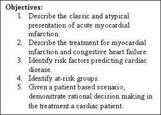
The following is an excerpt from my Clinical Decision Making lecture series on cardiac emergencies. In this selection I focus on the “nuts and bolts” of chest pain and congestive heart failure. The continuation module; SYNCOPE: The EMS Fatal Masquerade will be published shortly.

Previous Clinical Decision Making lectures include LOCATE the Patient and The Dead: Clinical Decision Making in Cardiac Arrest. The latter are under revision and will be re-issued in January 2006.

Cardiac Events and Congestive Heart Failure
It comes as no surprise to the experienced EMT or paramedic that a majority of patients requesting emergency medical service have complaints of cardiac or respiratory nature. These complaints are often categorized from the acute complaint of chest pain, syncope (or near-syncope), or short of breath to cardiac arrest. Most often, when someone calls EMS, there is little question as to what the problem is…especially when the chief complaint is stated as above. Difficulties arise when the chief complaint is less evident and patient presentation is less than acute as in the case of the “weak and dizzy”, the elder person who is just “not acting normal”, and the cold/flu patient.

The difficulty is for the provider to differentiate between the two and treat appropriately. Making the call between myocardial infarction, congestive heart failure (CHF), chronic obstructive pulmonary disease (COPD), and cold/flu can be difficult in any setting. Mistaking the patient in CHF for one who has respiratory infection can be deadly. Equally so, is the patient with syncope or near-syncope that claims to be “fine” now. Conducting a through assessment is critical to discovering what type of condition the patient may have, formulating a differential diagnosis list, and making treatment/transport decisions.

Chest Pain
Discovering the etiology or cause of chest pain can be difficult for EMS providers mainly because the pain is described in vague terms. Patients with cardiac related pain or discomfort often have visceral pain. A patient with visceral pain may complain that the symptoms are located in a general area and intermittent yet, worsen with time. They also note the nature of the pain as dull, aching, or numbness. By contrast, somatic pain is able to be localized to a specific area by the patient. This somatic pain may change with movement or position of the patient. A word of caution is due here; any pain in the chest, either somatic or visceral, must be taken seriously as a potential cardiac event. Fundamental BLS care including oxygen and evaluation by advanced life support is necessary.
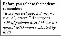
In addition to the quality (visceral or somatic) of pain, the location of chest pain is also non-diagnostic. That is; the location of pain often will not directly relate to the origin of the pain. Many groups of patients may not experience the “classic” symptoms of chest pain, shortness of breath (SOB), arm/neck pain, nausea, diaphoresis, and the like. Diabetics, elderly, and post-menopausal women may experience altered or atypical presentation of a cardiac event. Persons who are chronic drug abusers may also not report textbook symptoms. In any case, decreased severity or absence of pain does not equate to a non-life threatening condition in the setting of chest pain. In addition to the classic signs and symptoms of cardiac events, be on the lookout for the following list in addition to the classic symptoms:
· Weakness and fatigue
· Pressure in the chest
· Palpitations, or racing heart
· SOB or dypsnea…even if mild
· Painful digestion, dydpepsia
· Neck, back and epigastric pain
· Symptoms that wake the patient from sleep or keep them from sleeping
· Any change in mental status, syncope or near-syncope…even if the patient “is fine now”.

Any one of the above should indicate potential for a cardiac event, with or without chest pain. With these complaints, the EMT must consider acute myocardial infraction (AMI), angina (stable and unstable), cardiac dysrhythmias, and pulmonary embolism. Again, solid BLS care should be initiated as well as ALS assessment and transport.

Congestive Heart Failure
Congestive heart failure or CHF is a situation when one or both (univentricular and biventricular) of the heart ventricles is no longer capable of maintaining discharge capacity of fluid returning to the heart. The results may be seen in the form of any number of symptoms depending upon what side of the heart is failing.

This is a good time to remember some basic physiology of the heart. The right side of the heart is under low pressure in relation to the left heart as it receives blood from the body (pre-load) and is responsible for pumping into the pulmonary system for gas exchange. With this in mind, one can visualize that any factor the restricts or diminishes the right heart ability to push blood will result in a backup of fluid into the systemic circulation; the liver and dependant areas for example. This situation is compounded by inability to get blood to the pulmonary system for oxygenation. When this occurs, not only is oxygenation impaired with resulting hypoxemia, but the left ventricle (and cardiac output) also becomes challenged. Simply put, the left heart cannot pump required volume if that volume is not delivered (no pre-load, no after-load). These situations can arise from AMI, valve failure (tricuspid, pulmonary, or both), and fluid depletion. Paradoxically, the most common cause of right heart failure is left heart failure a.k.a Cor Pulmonale.

A similar situation occurs if the left or high pressure side of the heart is involved. The left heart can be thought of as the high pressure side and is responsible for ejecting blood to the body via the aortic valve and the aorta. This is known as after-load. Anything that reduces the left ventricles efficiency can technically cause left heart failure. Think of it this way:
If volume entering the left atria are insufficient, there will be decreased discharge
If the mitral (bicuspid) valve fails either by stenosis or regurgitation (narrowing or allowing backward flow), the left ventricle (LV) either cannot fill or losses discharge pressure and volume back to the left atria.
If the aortic valve is stenosed or narrowed, the LV must work harder to discharge the needed volume and pressure.
If the LV is damaged from AMI and not able to accommodate the preload being sent

These bullets indicate that LV failure can come from any number of causes: AMI, valve failure, and prolonged high systemic pressures (high blood pressure). Regardless of the cause, the results of decreased cardiac output and backup into the pulmonary system (high pulmonary pressures and pulmonary edema) may be seen.

Common Sense Stuff
Heart failure is basically a theory of backed-up fluid that accumulates behind the chamber (or side) of the heart that is affected. Patients with severe left ventricular infarction often exhibit symptoms of respiratory distress and hypoxia as a result of impaired pumping ability (decreased after load) and pulmonary edema as the existing pre load backs up behind the left ventricle. The increased pulmonary pressure is passed on the right ventricle and may eventually cause it to become impaired. In the setting of right heart failure, either resulting from left heart failure or right ventricular AMI, pre load decreases (remember: the left heart can’t pump out what it doesn’t get) and fluid backs up behind the right heart and into the systemic circulation. This fluid accumulation becomes visible as ankle edema, edema in dependant areas of the body, and liver engorgement (congestive hepatomegaly).

Is It Acute or Chronic?
We may find any number of symptoms of heart failure presented to the EMS provider. Unfortunately, the early or mild symptoms may be confused for cold/flu symptoms…a dangerous clinical assessment mistake! The symptoms depend not only on the side of the heart affected, but the rate at which pathology develops. Look at it this way; if a patient suffers a massive MI, arrhythmia such as rapid atrial fibrillation/ventricular tachycardia, or valve failure, drastic reduction of cardiac output and/or pulmonary edema may follow. If the patient goes on to survive any of the above or if conditions progress over a long period of time, the patient may compensate. Compensatory mechanisms may include enlargement of the heart, and many form of neurohormonal changes. In this case frequency of EMS contact and ED visits often increased.